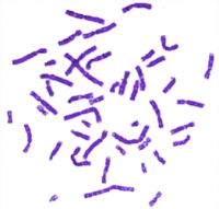


Giemsa stain is used in cytogenetics and for the histopathological diagnosis of malaria and other parasites.
Applications
It is specific for the phosphate groups of DNA and attaches itself to regions of DNA where there are high amounts of adenine-thymine bonding. Giemsa stain is used in Giemsa banding, commonly called G-banding, to stain chromosomes and often used to create a karyogram (chromosome map). It can identify chromosomal aberrations such as translocations and rearrangements.
It stains the trophozoite Trichomonas vaginalis, which presents with greenish discharge and motile cells on wet prep.
Giemsa stain is also a differential stain, such as when it is combined with Wright stain to form Wright-Giemsa stain. It can be used to study the adherence of pathogenic bacteria to human cells. It differentially stains human and bacterial cells purple and pink respectively. It can be used for histopathological diagnosis of malaria and some other spirochete and protozoan blood parasites. It is also used in Wolbach's tissue stain.
Giemsa stain is a classic blood film stain for peripheral blood smears and bone marrow specimens. Erythrocytes stain pink, platelets show a light pale pink, lymphocyte cytoplasm stains sky blue, monocyte cytoplasm stains pale blue, and leukocyte nuclear chromatin stains magenta. It is also used to visualize the classic "safety pin" shape in Yersinia pestis.
Giemsa stain is also used to visualize chromosomes.
Giemsa stains the fungus histoplasma, chlamydia bacteria, and can be used to identify mast cells.