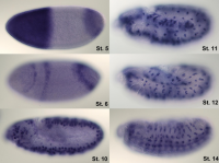


In Situ Hybridization (ISH) is a powerful technique that allows for specific localization of nucleic acid targets in fixed tissues, cells and circulating tumor cells (CTCs). The principle of ISH is that nucleic acids, when preserved adequately in a histologic specimen and when fixed appropriately, can be detected by using a nucleic acid probe, allowing you to obtain gene expression information in the context of tissue/cell morphology. In the past, quantitation of in situ gene expression, at the RNA (mRNAs or non-coding RNAs) level, had limited utility due to low sensitivity (e.g. non-radioactive formats), long complicated workflows (e.g. radioactive formats) and/or the inability to simultaneously multiplex.
In situ hybridization was invented by Joseph G. Gall.His work included major contributions to studies of chromosome structure and function, nucleic acid metabolism, gene structure, and cellular organelles.
In situ hybridization is used to reveal the location of specific nucleic acid sequences on chromosomes or in tissues, a crucial step for understanding the organization, regulation, and function of genes. The key techniques currently in use include: in situ hybridization to mRNA with oligonucleotide and RNA probes (both radio-labelled and hapten-labelled); analysis with light and electron microscopes; whole mount in situ hybridization; double detection of RNAs and RNA plus protein; and fluorescent in situ hybridization to detect chromosomal sequences. DNA ISH can be used to determine the structure of chromosomes. Fluorescent DNA ISH (FISH) can, for example, be used in medical diagnostics to assess chromosomal integrity. RNA ISH (RNA in situ hybridization) is used to measure and localize RNAs (mRNAs, lncRNAs, and miRNAs) within tissue sections, cells, whole mounts, and circulating tumor cells (CTCs).